Magnetic Resonance Imaging (MRI) scan
Magnetic resonance imaging (MRI)
is a painless, non-invasive and safe diagnostic technique.
It does not use X-rays,
but rather the natural magnetic properties of the human body.



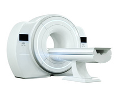
Find out how to prepare for your scan, what diagnostic equipment we use, and get answers to any questions you may have.
Magnetic resonance imaging (MRI)
is a painless, non-invasive and safe diagnostic technique.
It does not use X-rays,
but rather the natural magnetic properties of the human body.
This is a diagnostic procedure that uses a very strong electromagnetic field to produce detailed images of the anatomical structures of the human body. MRI produces images that enable the advanced diagnosis of a range of medical conditions, such as: cancers, developmental abnormalities, stroke, spinal injuries, joint diseases and dementia.
Magnetic resonance imaging is used, amongst other things, in situations where further diagnostic investigation is required and when the results of a CT scan are inconclusive. The indications for an MRI scan, either instead of or in addition to a CT scan, depend on the type of condition, the patient’s age and the diagnostic issue.

We recommend that you fast for 4 hours before the test (you may drink water)

The examination takes 15–60 minutes. During this time, you must remain still in one position.

People who take medication on a regular basis should take it as usual. People with diabetes must remember to take their insulin.

Please do not bring any metal objects into the MRI room, in particular mobile phones, watches, other electronic devices and credit cards.

Before the examination, please remove items such as: glasses, jewellery, watches, hearing aids and dentures.

Please arrive for your appointment 15 minutes before the scheduled time to complete the medical questionnaires.
For examinations requiring the administration of a contrast agent, patients are asked to drink at least 1.5 litres of fluids within 12 hours on the day before the examination.
We would ask anyone undergoing an MRI scan of the head to avoid wearing heavy make-up.
Pacemaker (particularly older models), insulin pump, cochlear implant, neurostimulators, intracranial metal clips, metallic foreign body in the eye

Metalowe endoprotezy, szwy i ciała obce w innej lokalizacji, pierwszy trymestr ciąży. O wykonaniu badania decyduje lekarz radiolog. W przypadku wątpliwości prosimy o kontakt telefoniczny z Kierownikiem Pracowni.

Remember!
If the examination is to be carried out under the National Health Fund (NFZ), the original referral must be submitted to the clinic 14 days before the scheduled date of the examination. This requirement applies only to paper referrals. It does not apply to e-referrals.
Please note that you can also register foronline survey.

The examination is painless and is carried out by a radiography technician
The examination takes
between 15 and 60 minutes

The patient lies comfortably, usually on their back

The test is accompanied by
increased noise levels.

During the examination,
you must not move

After the examination, you can return
to your daily routine
Please arrive for your scan 15 minutes before your appointment time to complete a medical questionnaire regarding any chronic conditions, allergies, medication you are taking and any possible contraindications to an MRI scan.
For some MRI scans, a contrast agent is administered, mainly intravenously. In such cases, a nurse will insert an intravenous cannula just before the scan begins. The contrast agents we use are very safe and rarely cause allergic reactions.
The patient is escorted into a special room by a member of the department’s staff and asked to lie down on a comfortable examination table, which is then slid into the scanner to scan the patient’s body.
Medical staff operate the scanner and monitor the scan from an adjoining room separated by a glass partition.
The scan is painless; although some patients may experience discomfort when the contrast agent is administered, this is not usually the case. A technician, a doctor and a nurse are on hand to look after the patient, and any instructions are given via a microphone.
Most MRI scans last between 15 and 45 minutes, depending on the area being examined and the number of images required, although sometimes a scan may last 60 minutes or longer. It is very important for the patient to remain still during the scan.
Following a scan involving the administration of a contrast agent, the patient is asked to remain in the scan suite for several dozen minutes as a precaution. Provided there are no adverse reactions, the patient may leave the premises.
The patient is asked to drink two litres of fluid a day for the next two days, as drinking plenty of fluids helps to ensure that the contrast medium is efficiently excreted from the body.
At reception, patients will be informed of the date on which they can collect their test results, which are sent to our specialists for interpretation and consultation. Results can be collected in person or by another person, provided the relevant authorisation form has been completed. The results consist of a medical report and a disc containing the MRI images, together with the report.
It is also possible to electronic receipt of results.
for the diagnosis of the brain, spinal cord and meninges in cases of vascular (stroke-related), metabolic, neoplastic and inflammatory conditions.
to accurately determine the location of a tumour within the head or spine, to plan surgical treatment, to distinguish between post-operative scarring and tumour recurrence, to locate the epileptic focus, to identify vascular malformations prior to surgical treatment, and to determine the cause of sciatica or other radicular syndromes prior to surgical treatment.
to assess the heart’s function and capacity.
for the diagnosis of diseases of the liver and bile ducts, diseases of the pancreas, the small and large intestines (including the rectum), diseases of the urinary tract and the prostate, for the diagnosis of perianal fistulas, and for the assessment of the foetus and the pregnant uterus.
to accurately assess the condition of the cartilage, ligaments and tendons in the shoulder, knee, hip, elbow, wrist and other joints. Where justified, an orthopaedic surgeon may refer a patient for an MRI scan of the joint with intra-articular administration of a contrast agent
to assess the condition of the middle and inner ear, in the diagnosis of tinnitus, vertigo and hearing loss, to assess the stage of laryngeal cancer, and to assess benign and malignant tumours of the neck, sinuses and salivary glands.
for the accurate assessment of diseases of the eyeball, the orbit and the visual pathways.
for the diagnosis of diseases of the pituitary gland, the adrenal glands and the thyroid gland.
MRI scans in children require appropriate preparation of the child before the scan, supervision during the scan and aftercare once it is complete. The specific nature of this type of scan means that they are carried out in specialised centres equipped with the appropriate staff, equipment and technical facilities.
Very few children are able to lie still for more than 30 minutes, which is why, in the vast majority of examinations involving children, the assistance of an anaesthetist is essential; their role is to put the child to sleep, thereby enabling the examination to be carried out whilst the child remains still.
For up to two hours after the examination, the child remains in the observation room under the care of staff until they are fully awake.
Please contact the registration desk on 32 701 24 00 in order to find the nearest clinic equipped to examine children.
You will find the eWUŚ declaration in the file below
In the file below, you will find the eWUŚ declaration to be used in cases where a patient has a legal guardian or a statutory representative


