Computed Tomography (CT) scan
Computed tomography (CT)



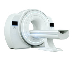
Find out how to prepare for your scan, what diagnostic equipment we use, and get answers to any questions you may have.
Computed tomography (CT)
It is a diagnostic imaging method in which multi-slice cross-sections of the patient’s body are obtained using X-rays. Owing to its extremely versatile range of applications, computed tomography has become the primary imaging method across all medical specialities. It is used, amongst other things, for imaging injuries, in examinations of the head, chest and abdomen, for imaging internal organs including the liver, pancreas and kidneys, and for detecting neoplastic lesions, haemorrhages and embolisms. CT is also used for virtual colonoscopy and non-invasive coronary angiography, as well as other angiographic examinations.

We recommend that you fast for 4 hours before the test (you may drink water)

The test takes a few minutes. During this time, you must remain still in one position.

People who take medication on a regular basis should take it as usual. People with diabetes must remember to take their insulin.

For examinations requiring the administration of a contrast agent, patients are asked to drink at least 1.5 litres of fluids within 12 hours on the day before the examination.

f there are any items in the vicinity of the part of the body being examined that could interfere with the quality of the diagnostic image, these must be removed before the examination. These include, for example: glasses, jewellery, a watch, a hearing aid and dentures.

Please arrive for your appointment 15 minutes before the scheduled time to complete the medical questionnaires

Remember!
If the examination is to be carried out under the National Health Fund (NFZ), the original referral must be submitted to the clinic 14 days before the scheduled date of the examination. This requirement applies only to paper referrals. It does not apply to e-referrals.
Please note that you can also register for online survey.

The examination is painless and is carried out by a radiography technician
The examination takes
a few/a dozen or so minutes

The patient lies comfortably, usually on their back

The test is not accompanied by
increased noise levels

During the examination,
you must not move

After the examination, you can return
to your daily routine
Please arrive for your scan 15 minutes before your appointment time to complete a medical questionnaire regarding any chronic conditions, allergies, medication you are taking and any possible contraindications to undergoing a CT scan.
For certain CT scans (e.g. of the abdomen and pelvis), a contrast agent is administered to obtain a clear image of the area being examined. Both intravenous and oral contrast agents are used, as well as intraintestinal contrast agents administered as infusions. The contrast agents we use are very safe and rarely cause allergic reactions. If oral administration of the contrast medium is required, please arrive at least one to two hours before your scheduled appointment and bring 1.5–2.5 litres of still mineral water with you. A technician or nurse will prepare the contrast solution, which you should then drink slowly in small sips. If the contrast is to be administered intravenously just before the examination begins, a nurse will insert an intravenous cannula.
Most CT scans follow a similar procedure. The patient lies down on a special movable table and remains in a lying position throughout the scan, usually on their back (or, alternatively, on their side or stomach), whilst the table moves through the scanner ring.
The scan is painless; although some patients may experience discomfort when the contrast agent is administered, this is not usually the case. A technician, a doctor and a nurse are on hand to look after the patient, and instructions are given via a microphone from an adjoining room separated by a glass partition.
During the scan, the patient may be asked to hold their breath and remain still, as any movement of the body may cause errors in the images obtained, which could result in a reduction in the quality of the results. The scan usually lasts from a few to a dozen or so minutes.
Following a scan involving the administration of a contrast agent, the patient is asked to remain in the scan suite for several dozen minutes as a precaution. Provided there are no adverse reactions, the patient may leave the premises.
The patient is asked to drink two litres of fluids a day for the next two days, as drinking plenty of fluids helps to ensure that the contrast medium is efficiently excreted from the body.
At reception, patients will be informed of the date on which they can collect their test results, which are sent to our specialists for interpretation and consultation. Results can be collected in person or by another person, provided the relevant authorisation form has been completed. The results consist of a medical report and a disc containing the CT scan images, together with a description.
It is also possible to elektroniczny odbiór wyników.
to determine the cause of headaches and dizziness, to rule out the presence of vascular malformations, brain and spinal cord tumours, to diagnose degenerative brain diseases, including dementia, to diagnose the cause of back pain, and to investigate other symptoms of neurological disorders.
to diagnose suspected brain and spinal tumours, and to identify the presence of vascular malformations, post-traumatic lesions and neoplastic lesions of the skull and spine.
to diagnose suspected brain and spinal tumours, and to identify the presence of vascular malformations, post-traumatic lesions and neoplastic lesions of the skull and spine.
for the diagnosis of pathologies of the lungs and chest wall in both benign and malignant conditions, and for the assessment of mediastinal lymph nodes, diagnosing disorders of the oesophagus, pulmonary embolism, pneumoconiosis and pulmonary fibrosis, and assessing the liver, bile ducts, pancreas, stomach, small intestine and large intestine.
for the diagnosis of disorders of the kidneys, ureters, bladder, prostate, uterus and ovaries.
to assess the pituitary gland in patients for whom there are contraindications to an MRI scan of the pituitary gland or the adrenal glands.
to assess the musculoskeletal system following injury and surgery, and prior to planned surgery, as well as in the diagnosis of tumours, degenerative diseases, osteitis, Paget’s disease and fibrous bone dysplasia.
to locate and determine the extent of the cancer, as well as prior to planned treatment, during treatment, and to monitor the effects of treatment.
to diagnose acute and chronic surgical conditions of the abdomen and pelvis, and following injuries to the torso or chest. A CT scan is often performed to assess the condition of major blood vessels, such as the aorta, prior to planned surgery.
to determine the nature of nodular lesions of the neck, including those of the larynx, and of diseases of the paranasal sinuses, the nasal cavity, the throat, the ears and the salivary glands.
in order to improve the diagnosis of inflammatory, neoplastic and post-traumatic changes in the orbit.
It is intended for patients who meet the following criteria:

aged between 50 and 75

long-term smoking

a family history of lung cancer and/or head and neck cancers
You will find the eWUŚ declaration in the file below
In the file below, you will find the eWUŚ declaration to be used in cases where a patient has a legal guardian or a statutory representative


