


SPECT scans
A modern diagnostic method
A modern diagnostic method
SPECT scans are used in the diagnosis of diseases of the bones, kidneys, nervous system, heart and blood vessels, thyroid and liver. However, it is worth mentioning other groups of conditions in which isotope scans are also used:
Myocardial perfusion scintigraphy (cardiac SPECT) is a non-invasive stress imaging technique, used primarily in patients with ischaemic heart disease.
The method is highly useful in the diagnosis of coronary artery disease and in risk stratification in patients with diagnosed coronary artery disease – as a screening test for coronary angiography and coronary revascularisation.
Indications for testing in accordance with the ESC/PTK guidelines on the management of stable coronary artery disease (2013) and coronary revascularisation (2018).
The examination involves assessing the distribution of the radiopharmaceutical 99mTc-MIBI – a substance labelled with a radioactive isotope – within the myocardium. The 99mTc-MIBI (methoxyisobutylisonitrile) tracer accumulates physiologically in the mitochondria of cells in a normally perfused heart via passive diffusion, in proportion to local blood flow in the coronary arteries. Ischaemia impairs this process and results in reduced uptake of the tracer by the mitochondria of cardiomyocytes. A visual representation of this process is obtained using a gamma camera.
The ability to assess the location and extent of ischaemia using cardiac SPECT is useful for risk stratification, thereby helping to avoid unnecessary invasive procedures.
Cardiac scintigraphy (cardiac SPECT) is a diagnostic test used to assess suitability for coronary angiography and coronary revascularisation.
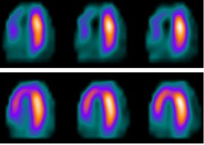
The procedure consists of two stages: a resting scan and a stress scan. Depending on your preference, both scans can be carried out on the same day or over the course of two visits to the nuclear medicine department. The total duration of the examination is a maximum of 5 hours. The stress test involves either physiological exercise on a treadmill or pharmacological stress – the latter is used in patients who, for whatever reason, are unable to perform adequate physical exercise, as well as in patients with an uninterpretable ECG recording: LBBB, or a rhythm generated by an implantable device, such as an ICD.
The patient receives the radiopharmaceutical intravenously on two occasions: at rest and during exercise or following administration of a medication. Images are then recorded using a gamma camera in a tomographic mode, producing maps of radioactivity distribution in the heart muscle.
The interpretation of the test involves comparing the images obtained in both phases. Normal coronary flow is characterised by uniform tracer accumulation both at rest and during exercise. A reduction in 99mTc-MIBI accumulation following exercise, in conjunction with a normal resting scan result, indicates reversible ischaemia (Fig. 1). Reduced tracer uptake in the same area at rest and after exercise indicates the presence of scarring.
The negative predictive value of cardiac SPECT is very high and, according to the literature, is as high as 98.8 per cent, which means that a negative result rules out the possibility of a heart attack and/or cardiac death in 98.8 per cent of patients who underwent the scan during an average follow-up period of 2 to 5 years.
Cardiac SPECT is the preferred method over an electrocardiographic stress test in patients who have previously undergone coronary intervention: PCI or CABG.
Compared with a conventional exercise ECG, the method of isotope-based myocardial perfusion imaging is characterised by significantly higher sensitivity: it offers better diagnostic performance in terms of detecting CAD with significant stenoses, providing a quantitative assessment and localisation of areas of ischaemia, and the ability to make a diagnosis in cases where abnormalities are present on a resting ECG: e.g. LBBB or pacemaker rhythm, where SPECT is the method of choice.
Pregnancy, a previously known allergy to MIBI. Renal failure is not a contraindication for isotope scanning.
The presence of a large area of reversible myocardial ischaemia (>10%) is associated with a poorer prognosis and helps to identify patients in whom further diagnostic investigations, including coronary angiography, should be carried out and revascularisation performed. The percentage figures are always stated on the report.
A specialist doctor – a cardiologist or a doctor of another speciality – may refer a patient for a myocardial perfusion scan. The patient attends the appointment with their referral and cardiology records.
The examination is reimbursed if the referrals are issued by a specialist clinic with a current contract with the National Health Fund (NFZ) or by a hospital ward on the day of the patient’s discharge: the discharge summary must include the statement ‘a referral for a cardiac SPECT scan has been issued’, and the patient must be provided with the referral in paper or electronic form. The referring institution does not bear the cost of the examination.

Please note
Two referrals to the Department of Nuclear Medicine are required: one for a resting scan and one for a stress scan (exercise or pharmacological test).

ul. Ceglana 35
40-514 Katowice
Mon–Fri: 7:00 – 21:00
