


- Select a test
Patient Guide
Find out how to prepare for your scan, what diagnostic equipment we use, and get answers to any questions you may have.
Find out how to prepare for your scan, what diagnostic equipment we use, and get answers to any questions you may have.
A prostate biopsy is used to detect prostate cancer. It is carried out on men to confirm or rule out a suspected case of cancer. The procedure involves taking a sample of prostate tissue for histopathological examination. Treatment for prostate cancer can only be started on the basis of the results of a histopathological (microscopic) examination. There is no other test that can confirm the presence of this cancer.
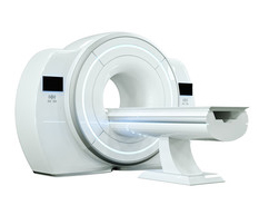
At Voxel Medical Diagnostic Centres in Warsaw, we perform precise prostate biopsies using real-time image fusion of multiparametric magnetic resonance imaging (mpMRI) and ultrasound. We have the technology to perform the procedure via both the transrectal and transperineal routes. All biopsies are carried out under anaesthesia.
Prostate cancer is the second most common cancer in men, after lung cancer. According to available data, the incidence of prostate cancer in Poland is estimated at over 12,000 cases a year. This accounts for 15.5 per cent of all malignant tumours in men.
The incidence of prostate cancer in Poland is around 45 per cent lower than the average for European Union countries. However, despite this, mortality rates in Poland are in line with the European average. This puzzling disparity is due, amongst other things, to the late diagnosis of prostate cancer and indicates that Polish patients have poorer prospects of recovery.
The conventional prostate biopsy has many limitations. The main one is the difficulty in visualising lesions on transrectal ultrasound. Around 70% of these lesions are not visible on ultrasound. In most cases, therefore, the conventional prostate biopsy remains a so-called systematic (mapping) biopsy, i.e. a ‘blind’ search for areas of malignant growth (cancer) by taking samples (usually 10–12) from various sites within the gland. The reliability of a biopsy performed in this way remains questionable. It is estimated that the initial biopsy misses around 21–47% of prostate cancers.
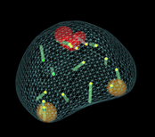
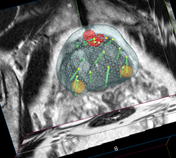
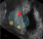
The solution to this problem is the early detection of prostate cancer via mpMR imaging and the replacement of routine biopsy with targeted biopsy. The technique that allows magnetic resonance images, which precisely show the location of the cancer, to be superimposed onto real-time ultrasound images is known as image fusion. From the patient’s perspective, the fusion biopsy procedure itself is very similar to a conventional biopsy. However, it allows for much more precise sampling of tissue from affected areas, whilst reducing the number of ‘missed’ samples.
In 2015, the European Association of Urology revised its guidelines on how to perform a prostate biopsy. It is recommended that any repeat prostate biopsy be carried out on the basis of an mpMRI scan and any abnormalities identified therein. As the method develops and economic conditions improve, it is to be expected that initial biopsies will also be carried out in this way.
Compared with mapping (cognitive) biopsy, fusion biopsy offers the highest probability of obtaining a tissue sample from the correct location for histopathological examination.
At the Voxel Medical Centre in Warsaw, precise prostate biopsies are performed using both the transrectal and transperineal methods. All biopsies are carried out under anaesthesia, the extent of which depends on the invasiveness of the procedure, medical indications and the patient’s decision.
Although not all patients undergoing mpMR and mpMR/ultrasound fusion biopsy procedures benefit from them, the following groups of patients are most likely to benefit:
An mpMR scan allows for a more accurate assessment of whether the neurovascular bundles are involved and, consequently, whether a nerve-sparing prostatectomy is possible. Furthermore, the mpMR scan enables the precise determination of the tumour’s stage (staging).
Such a situation can be frustrating for both the patient and the doctor. Thanks to mpMR/ultrasound fusion biopsy, a tool is now available that can significantly reduce the number of biopsies required to confirm the presence of prostate cancer
Thanks to mpMR/ultrasound-guided fusion biopsy, it is possible to determine more accurately whether the right patients are being monitored and whether any clinically significant cases of prostate cancer are being missed
The procedure for preparing for a biopsy is determined on a case-by-case basis by the urologist performing the biopsy, in consultation with the patient. There is no single, universal protocol for preparing the patient; rather, it is a set of guidelines that are applied depending on the patient’s condition, age, any underlying medical conditions and the medication they are taking.
A prostate biopsy is an invasive procedure, i.e. it breaks the continuity of the tissues. It therefore requires proper preparation:
A biopsy is performed by puncturing the prostate and removing a sample of glandular tissue for histopathological examination. The most common method of puncture involves accessing the prostate via the rectum or the perineum. This is carried out under ultrasound guidance.
In the case of a transrectal biopsy, the urologist inserts an anaesthetic gel and an ultrasound probe through the rectum to assess the prostate. Before the introduction of fusion biopsy, samples were taken from 12 typical sites, in the hope of ‘hitting’ the lesion by chance. However, the current fusion biopsy technique, using mpMR images, allows for the targeted collection of samples from sites visible and marked by the radiologist on the images of the lesions.
During the biopsy, the urologist has mpMR images with the lesions highlighted displayed on their monitor. These images are then merged/overlaid in real time with the ultrasound images. This allows, under ultrasound guidance, for targeted biopsies to be taken from sites visible on the mpMR images as areas of focal lesions. After the targeted biopsies have been taken, 12 standard sites are also biopsied. If no suspicious focal lesions are identified on the mpMR images that could serve as targets for biopsy, samples are taken only from the aforementioned 12 standard sites.
A transrectal biopsy is the most commonly used and least complicated procedure. Before the examination, the doctor inserts an anaesthetic gel into the rectum, followed by the transrectal ultrasound (TRUS) probe. Attached to the probe is a biopsy attachment, through which the urologist inserts a TRU CUT needle. This is a specially designed needle used to collect tissue samples 10–20 mm long and approximately 1 mm in diameter. Whilst performing the TRUS, the doctor determines the number of samples required and takes core samples from the prostate gland. These are then assessed by a specialist histopathologist, who makes the final diagnosis based on microscopic examination.
This procedure differs in the route of access to the prostate, which runs through the area of skin between the scrotum and the rectum. This technique is more complex and is therefore usually reserved for specific situations. Due to the nature of the procedure, a transperineal biopsy is carried out under general anaesthesia with the assistance of an anaesthetist, who ensures the patient’s complete comfort during the procedure.
The benefits of using this route of administration include a lower risk of infection, which, in an era of growing antibiotic resistance among gut bacteria, is of particular significance in:
Furthermore, as the needle travels along the peripheral zone, it provides easier access to the anterior portion and the apex of the prostate, which is particularly useful in patients who require a repeat biopsy.
A prostate biopsy is a relatively safe procedure, although complications can sometimes occur afterwards. For a few days following the procedure, minor, self-limiting bleeding may occur, which may manifest as blood in the semen (red or rust-coloured), in the urine or in the stools. In the vast majority of cases, these do not require medical intervention.
In the event of severe, persistent bleeding from the urinary tract or rectum, you should go to the nearest hospital without a referral. In less than 5% of patients, a genitourinary infection may occur despite the use of prophylactic antibiotics. If your temperature exceeds 38 ºC and/or you experience chills, you should go to the nearest hospital.
In around 0.2% of patients, temporary urinary retention may occur due to swelling of the prostate tissue following a biopsy. If you are unable to pass urine, you should go to the urology A&E department.


